Equipment
|
Image |
|
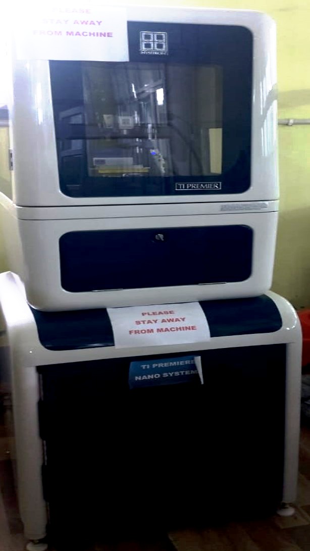
Nano Mechanical System
The TI Premier makes nanoscale mechanical and tribological characterization simple and consistent.Read more |
|
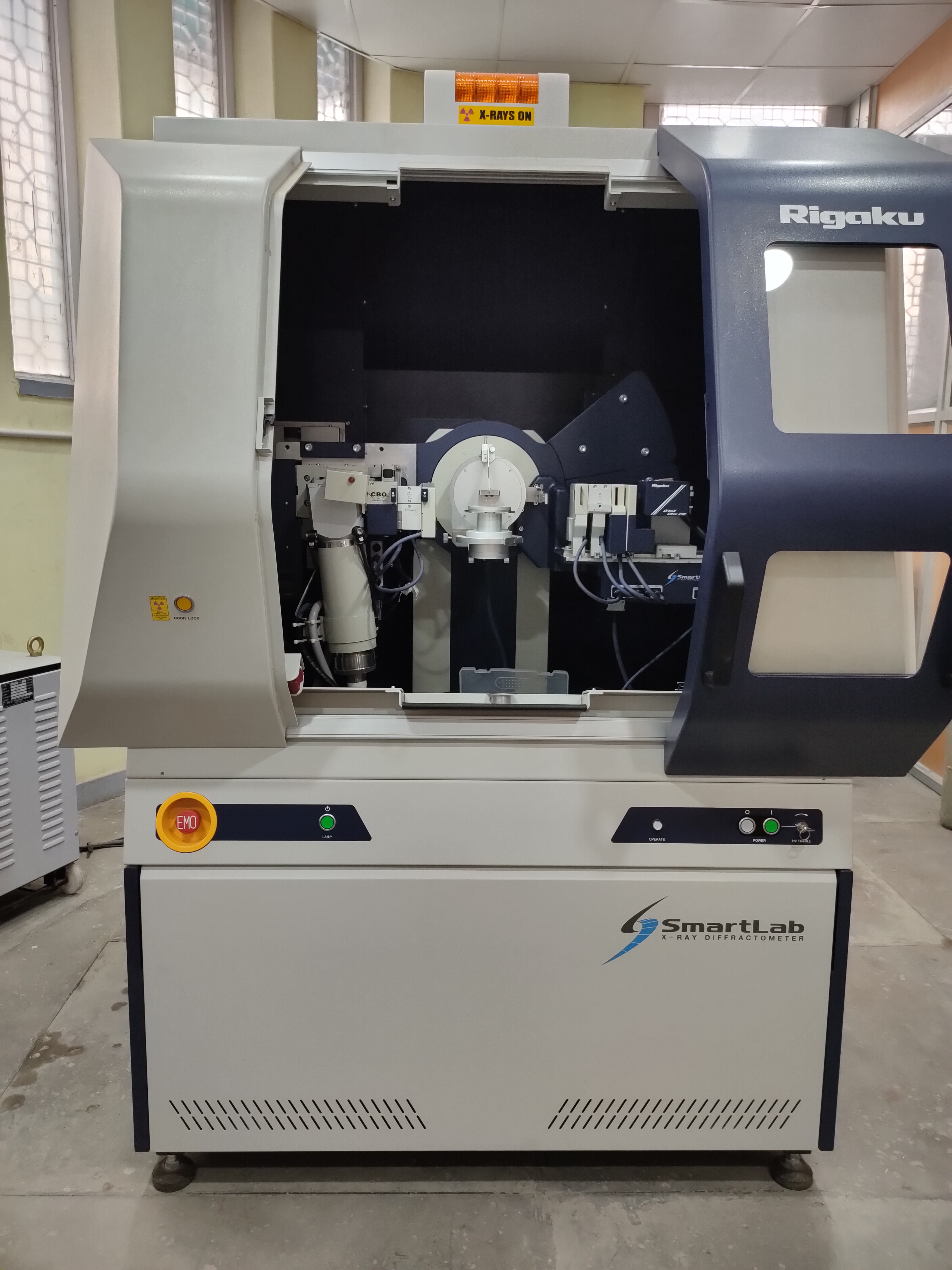
| SmartLab X-Ray Diffractometer (XRD)
The SmartLab 3kW XRD from Rigaku is a high-performance X-ray diffractometer that is designed for a wide range of applications, including powder diffraction, thin film metrology, small angle scattering (SAXS), in-plane scattering, and operando measurements. The system features a 3 kW rotating anode X-ray source. The SmartLab software provides the user with an intelligent User Guidance expert system functionality that guides the operator through the intricacies of each experiment.
Here are some of the key features of the SmartLab 3kW XRD:
- 3 kW rotating anode X-ray source for high-throughput and high-resolution measurements
- SmartLab Studio software with User Guidance expert system for easy operation and data analysis
The SmartLab 3kW XRD is a versatile and powerful X-ray diffractometer that is ideal for a wide range of applications in research and development, quality control, and materials science.
Here are some additional details about the SmartLab 3kW XRD:
- The 3 kW rotating anode X-ray source provides high-throughput and high-resolution measurements. This makes it ideal for applications where speed and accuracy are important, such as quality control and materials science research.
- The SmartLab Studio software with User Guidance expert system makes it easy to operate and analyze data. This makes it ideal for users with a range of experience levels, from beginners to experts.
Overall, the SmartLab 3kW XRD is a versatile and powerful X-ray diffractometer that is ideal for a wide range of applications in research and development, quality control, and materials science.
|
|
|
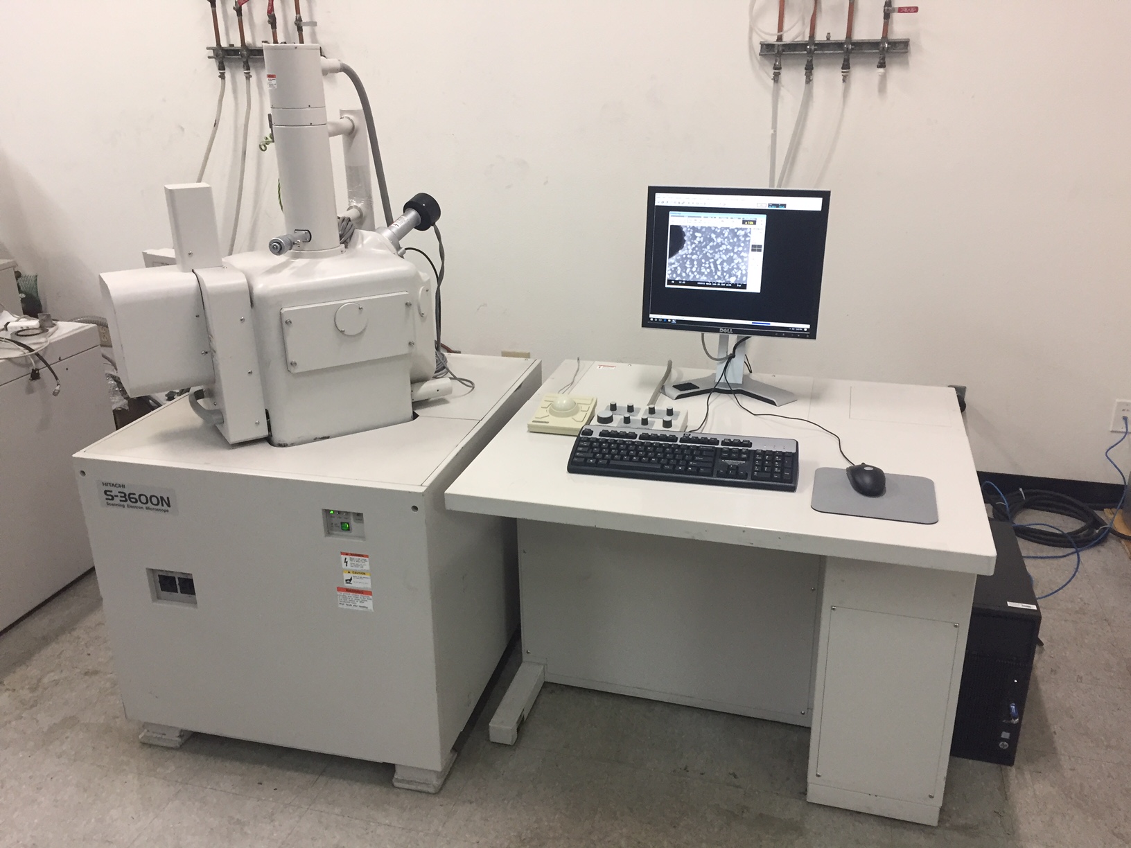
Scanning Elelctron Microscope
Hitachi 3600 N Scanning electron microscope with a 5 axis motorized stage coupled with ultra dry Compact EDS Detector Read more |
|
|
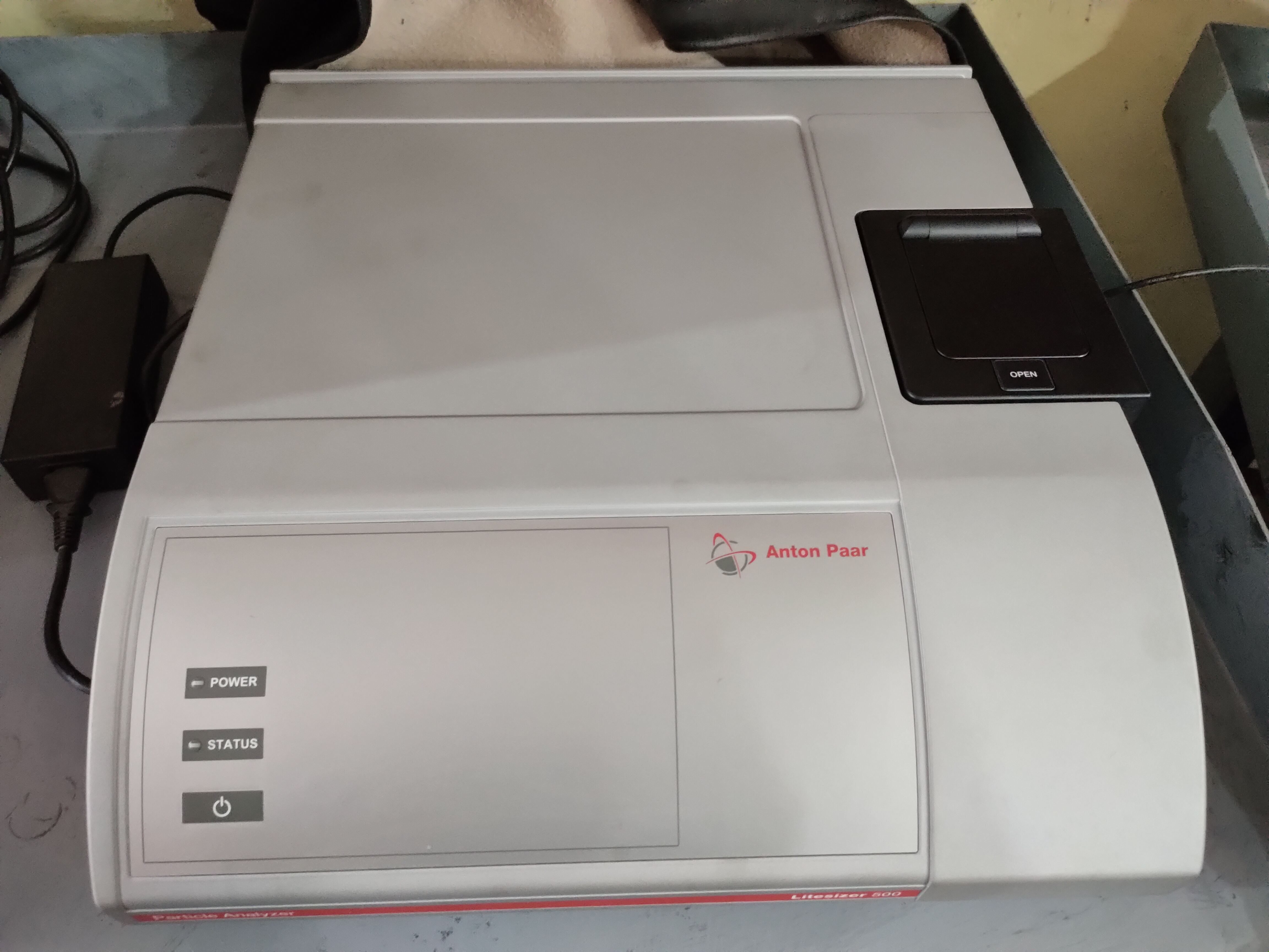
The Litesizer™ 500 Particle Size Analyzer
The Litesizer is an instrument for characterizing nano and microparticles in dispersions and solutions.
|
|
|
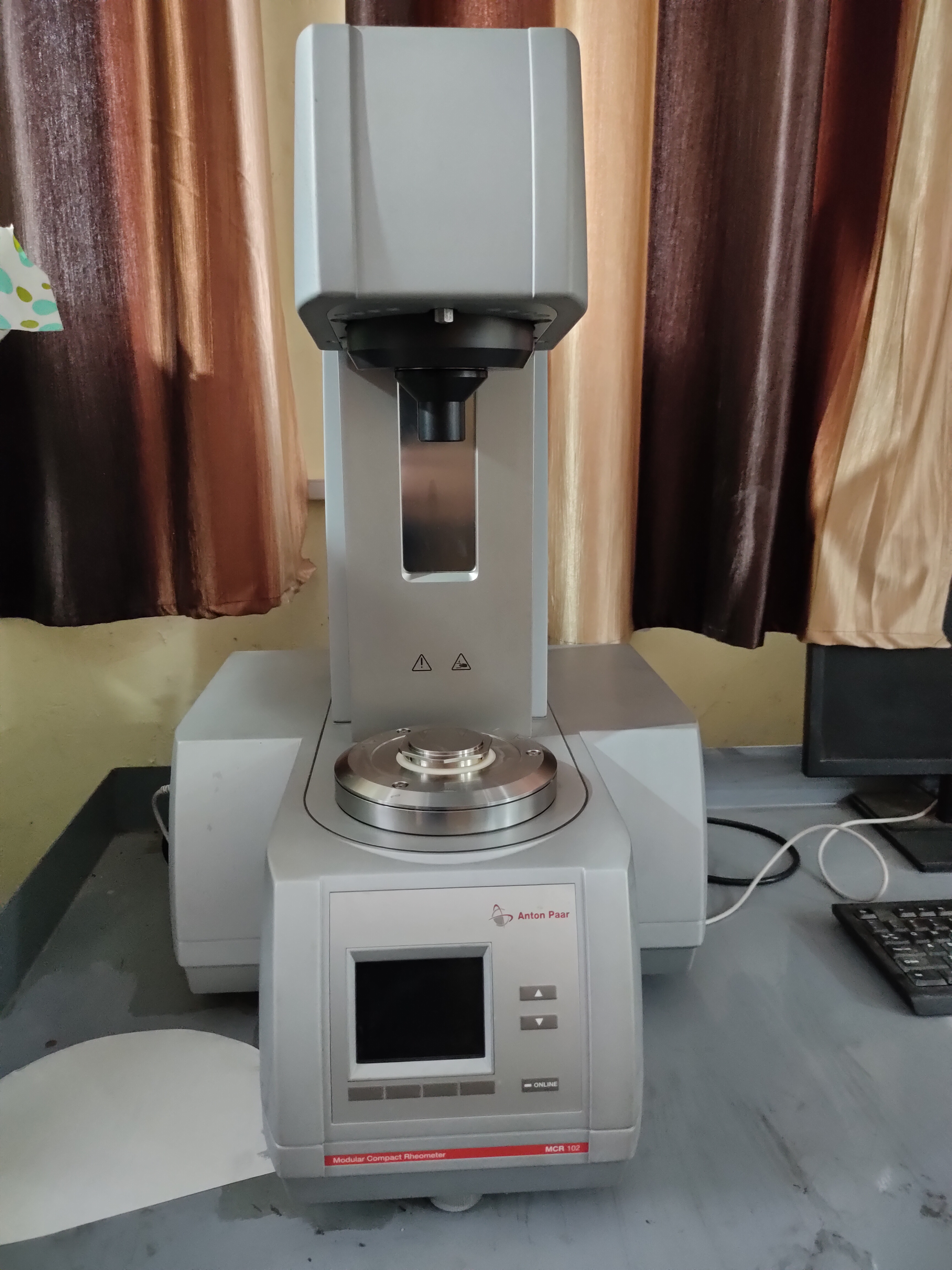
Anton Paar MCR 102 Rheometer
A Rheometer is a laboratory device used to measure the way in which a liquid, suspension or slurry flows in response to applied forces.
|
|
|
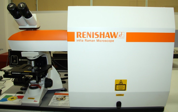
Raman Spectroscope
inVia Raman microscopes from Renishaw are high-sensitivity systems with integrated research grade microscopes, enabling high resolution confocal measurements. inVia Raman microscopes support multiple lasers, with automatic software switching of excitation wavelength
|
|
|
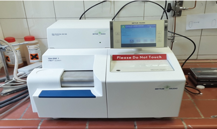
TGA/DSC
TGA/DSC measures the mass of the sample and energy absorbed as it is heated or cooled.Read more |
|
|
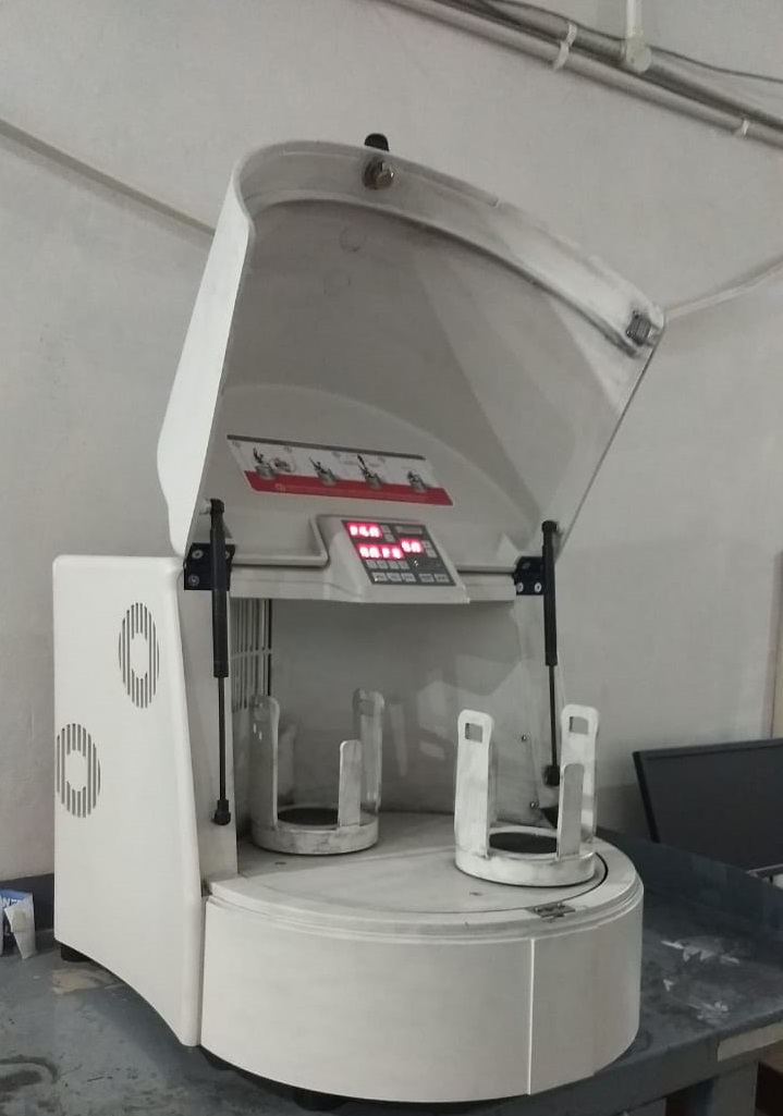
Planetary Ball Mill – “Mill pulverisette 5”
The laboratory planetary mill "pulverisette 5" can be used universally for high-speed grinding of liquid or solid inorganic and organic samples for analysis, quality control or material testing.Read more
|
|
|
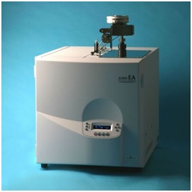
CHNS Analyzer
CHNS elemental analyzers provide a means for the rapid determination of carbon, hydrogen, nitrogen and sulphur in inorganic matrices and other types of materials.
|
|
|
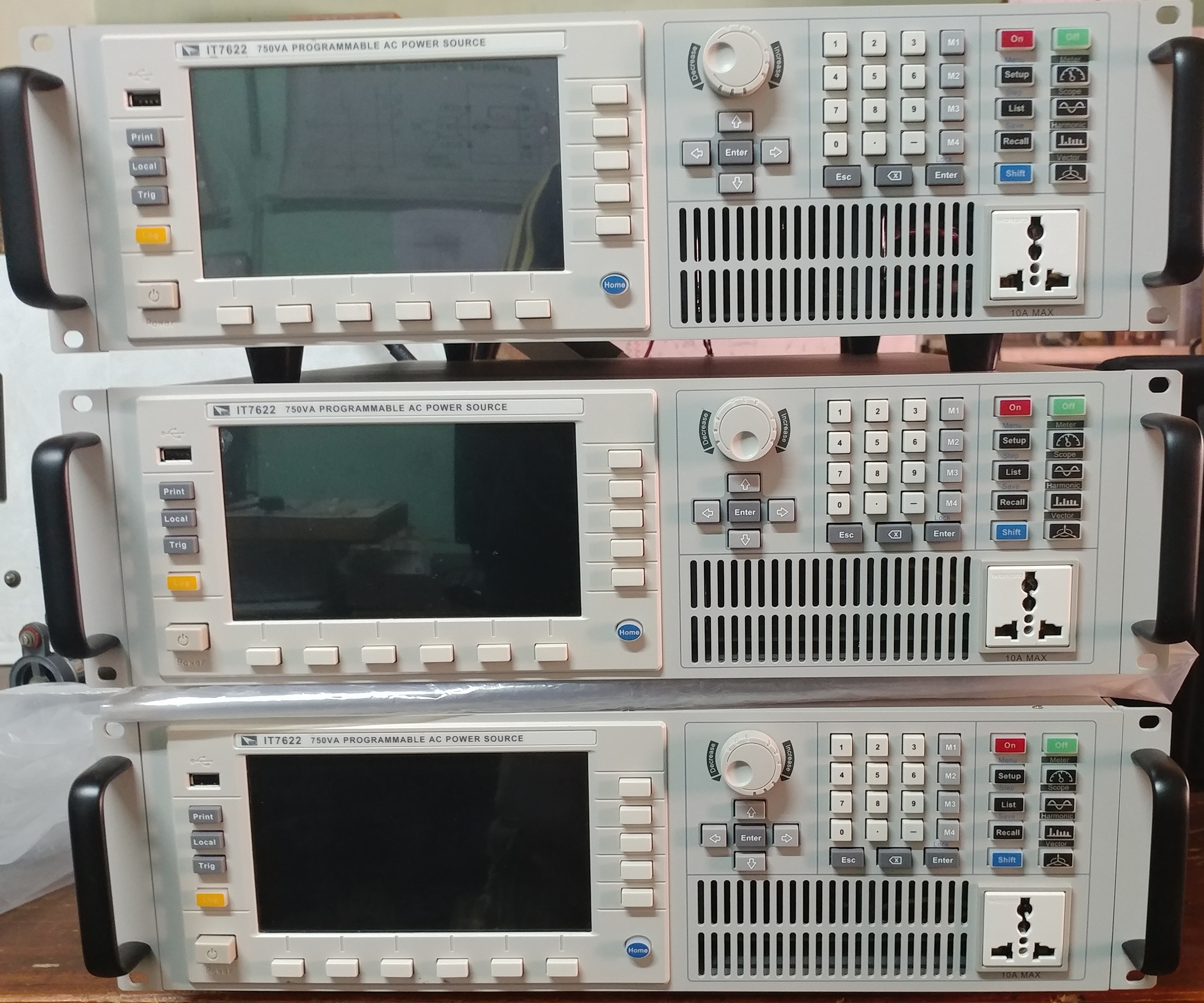
ITECH make 750VA AC Power Supply
Output frequency:10Hz-5kHz,Maximum Output voltage: 300 Vrms,Resolution 10 mV,RMS Current: 3 A at 300 V, 6 A at 150 V,Peak Current: 9 A at 300 V, 18 A at 150 V,More units can be combined to delivery higher current and 3-phase operation.
|
|
|
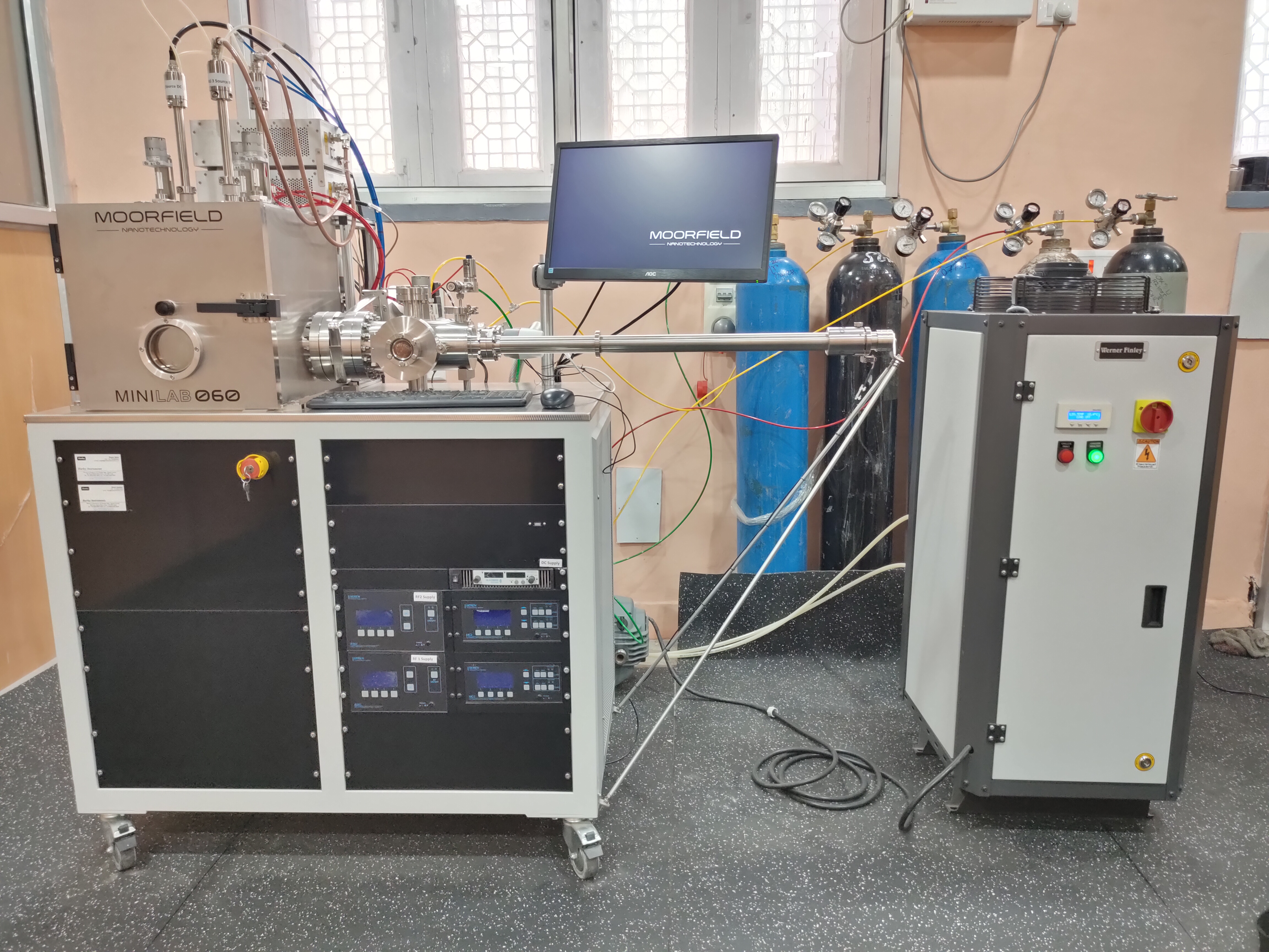
Multi Target Sputtering System
The MiniLab 060 standard configuration includes a turbo molecular pump positioned on an ISO160 port at the rear of the vacuum chamber. The vacuum chamber sits on a double-rack frame that contains all system control electronics and power supplies. MiniLab 060 system is available with load-lock facility.
|
 |
|
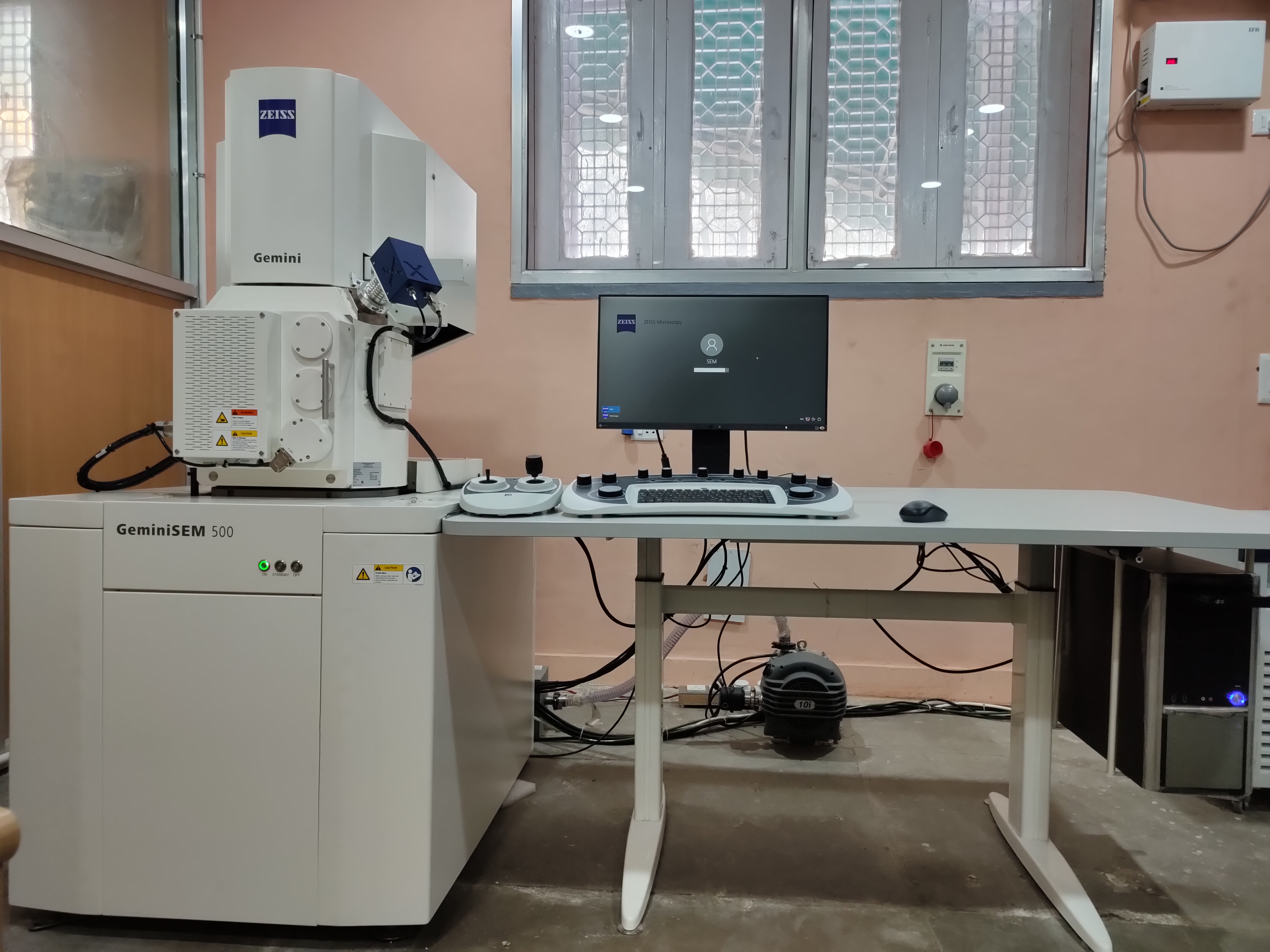
Field Emission Scanning Electron Microscope (FESEM)
The Gemini SEM 500 from Carl Zeiss is a high-resolution field emission scanning electron microscope (FE-SEM) that is used for a wide range of applications, including materials science, semiconductor failure analysis, and life sciences. The Gemini SEM 500 features a Nano-twin lens that enables rapid, reliable, and damage-free characterization of nanoscale defects and sensitive resist structures at low beam energies. The efficient detection allows you to operate at low currents for minimum beam damage, while enjoying excellent materials contrasts.
Here are some of the key features of the Gemini SEM 500:
- High resolution: The Gemini SEM 500 can achieve a resolution of 0.5 nm at 15 kV, 0.9 nm at 1 kV, and 1.0 nm at 500 V. This makes it ideal for imaging nanoscale features.
- Nano-twin lens: The Nano-twin lens is a unique feature of the Gemini SEM 500 that allows it to image beam-sensitive, nanoscale structures with minute detail at low beam energy.
- Efficient detection: The Gemini SEM 500 has an efficient detection system that allows it to operate at low currents for minimum beam damage. This results in better image quality and less sample damage.
- Wide range of applications: The Gemini SEM 500 is used for a wide range of applications, including materials science, semiconductor failure analysis, and life sciences.
The Gemini SEM 500 is a powerful tool that can be used to image and analyze a wide variety of materials. It is ideal for users who need high resolution and excellent image quality.
Here are some of the applications of the Gemini SEM 500:
- Materials science: The Gemini SEM 500 can be used to study the structure and properties of materials at the nanoscale. This information can be used to understand the behavior of materials and to develop new materials with improved properties.
- Semiconductor failure analysis: The Gemini SEM 500 can be used to image and analyze defects in semiconductor devices. This information can be used to identify the cause of failures and to improve the reliability of semiconductor devices.
- Life sciences: The Gemini SEM 500 can be used to image and analyze biological samples, such as cells and tissues. This information can be used to study the structure and function of cells and tissues and to develop new medical treatments.
|
|
|
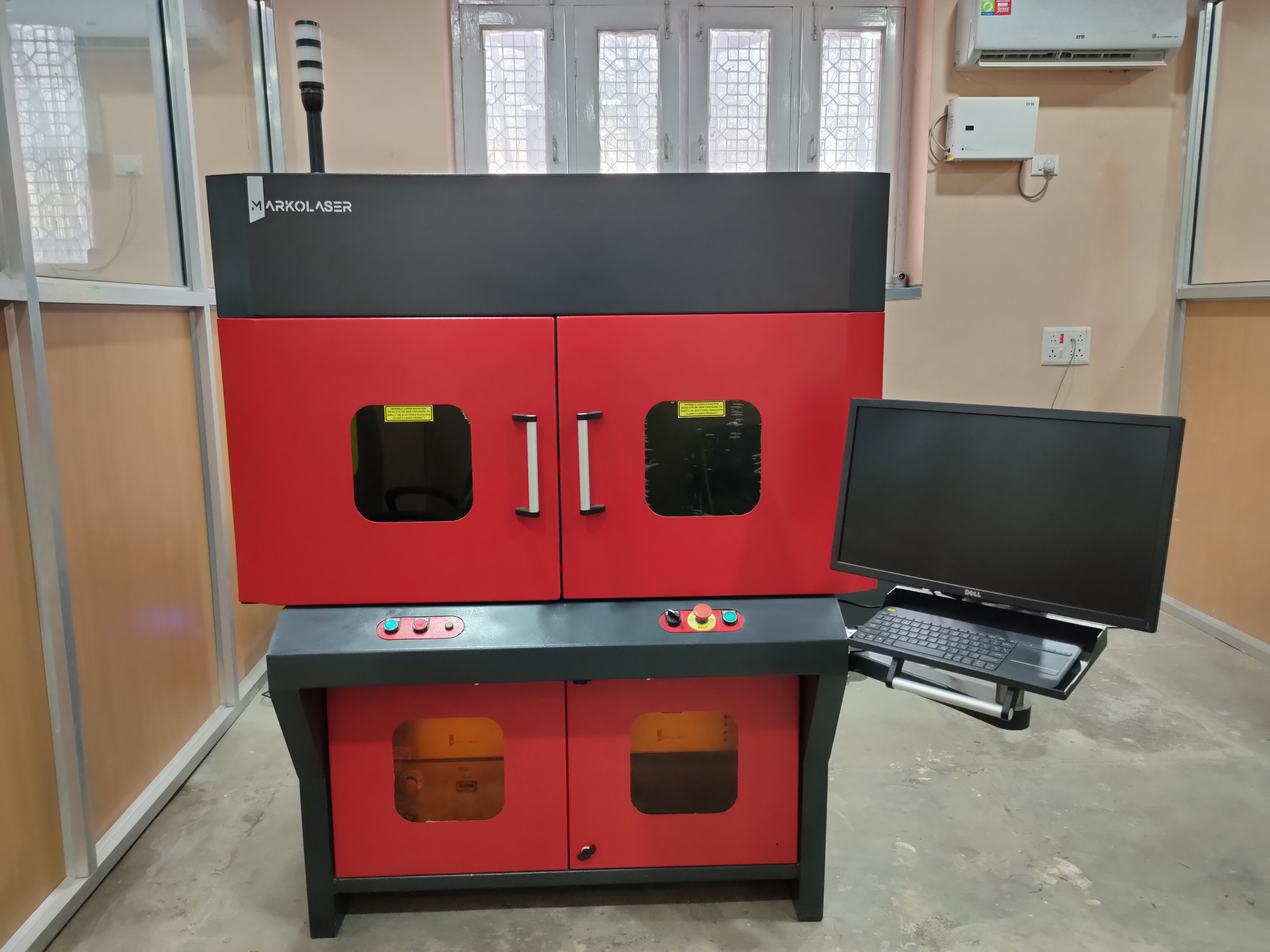
3-AXIS LASER SYSTEM FOR SURFACE TEXTURING
- Laser Unit, 3-Axis Laser System with Dual Heads, Machine Specifications, Electrical Power Requirement 110/220 VAC, Operating Temperature 10°C-35°C, Operating Humidity 5%-95%
- Cooling Air cooled, Marking Area 60 x 60 mm (For laser source 1) and 100x100 mm (For laser source 2), Marking preview gives the operator a chance to preview the pattern and adjust the component accordingly.
- Highly recommended for error-proof laser operation.
- Red pointer (useful to fine-tune laser focus)
- Useful when laser operations need to be performed on different components which differ in thickness & shape.
|
 |